
Body Composition 26 min read
Body Composition Analysis vs the Scale: 10 Key Differences
Your weight is a number. However, it is, in clinical terms, one of the least informative numbers you can have about your health. In fact, your weight tells…
Reading about body composition? Find an InBody test centre near you →
Your weight is a number. However, it is, in clinical terms, one of the least informative numbers you can have about your health. In fact, your weight tells you very little about your body. It does not show how much muscle you have. It also does not reveal where your fat is stored. More importantly, it cannot tell if your organs are surrounded by dangerous visceral fat. It also fails to show cell inflammation or early health risks. These include diabetes, heart disease, and sarcopenia.
As a result, many Indians receive normal-looking health reports every year. Their weight and BMI may appear fine. Their blood reports may also look borderline normal. However, hidden health risks may already be present. If left unchecked, these risks can contribute to serious illness over the years.
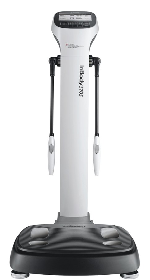
This is where precision body scanning becomes important. InBody’s Direct Segmental Multi-frequency Bioelectrical Impedance Analysis (DSM-BIA) technology goes beyond basic tools. It provides insights that a scale, BMI chart, or standard blood test cannot offer, showing what your body is truly made of in just 60 seconds. Published validation studies report close agreement between DSM-BIA and DEXA for lean mass and fat mass in adults, although BIA values are estimates and can differ from DEXA for an individual.[1][2][3]
Furthermore, it measures 10 different health parameters. Each one reflects a unique aspect of your metabolic health. In this article, we will explore all 10 parameters. We will also explain the clinical importance behind each one. This helps patients, clinicians, and health professionals understand what standard tests often miss.
| ICC 0.93–0.97 Agreement between DSM-BIA and DEXA for whole-body lean and fat mass in 484 middle-aged adults (published validation study)[1] | 10 Clinically distinct health markers revealed by a single 60-second InBody scan | 101M Indian adults estimated to be living with diabetes in the ICMR-INDIAB national survey (11.4% prevalence)[4] | 39.5% Of Indian adults have abdominal obesity, compared with 28.6% who have generalised (BMI-based) obesity (ICMR-INDIAB)[4] |
CLINICAL CONTEXT
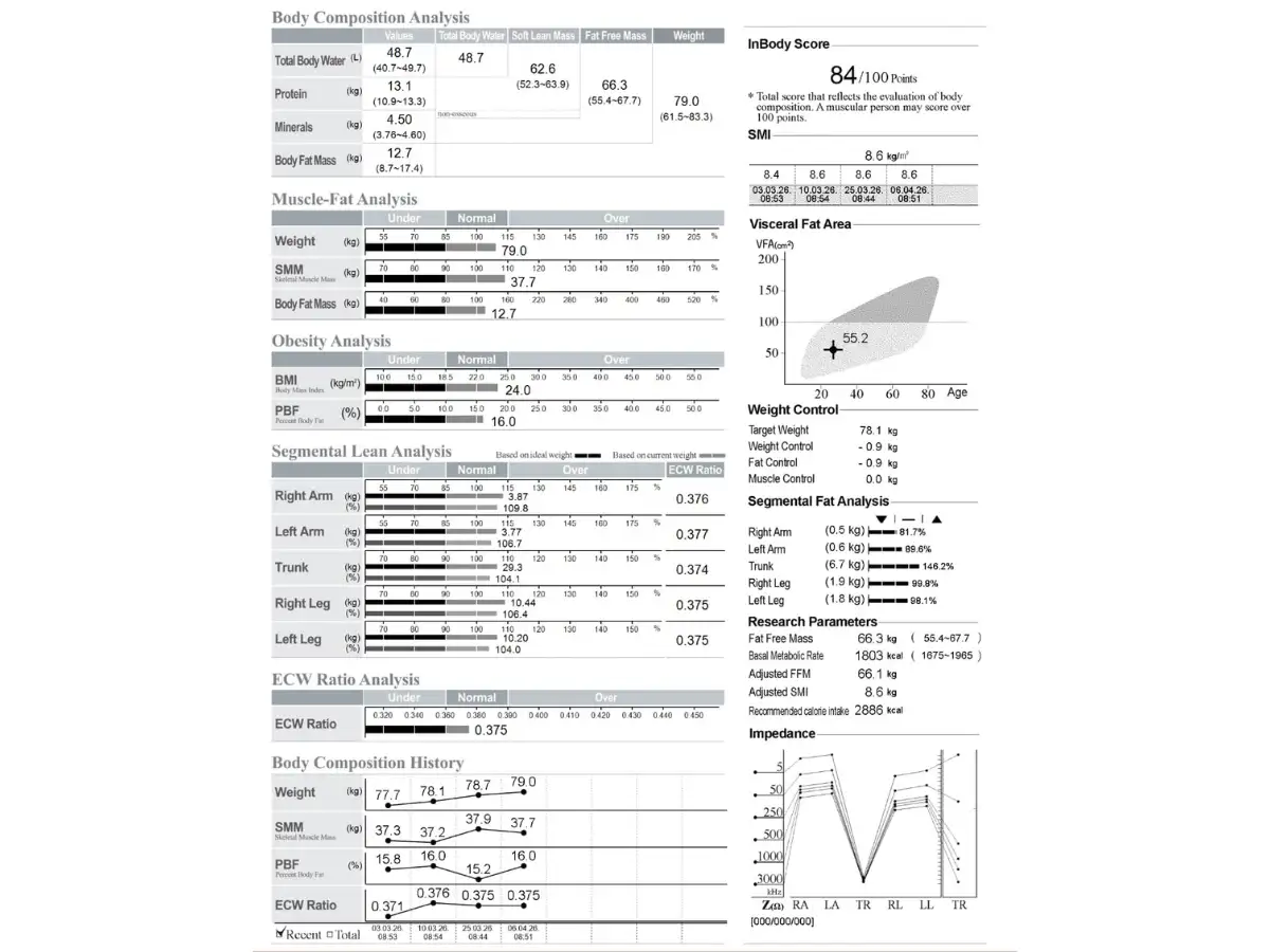
This article is written for both medical professionals and informed patients. The 10 precision body scan insights described below draw on published research, listed in the References at the end of this article. They are produced by InBody DSM-BIA technology, which has been compared with DEXA, a widely used reference method for body composition, in published validation studies.[1][2] Some outputs (such as BMR and segmental fat) are estimates calculated from the impedance data, and a scan supports, but does not replace, a clinical assessment.
Why Standard Health Screening Misses What Matters Most
Before examining what a precision body scan reveals, it is important to understand why standard health screening in India is structurally inadequate for detecting the metabolic risks that most commonly drive serious disease. Standard health checks in India typically include weight, BMI, blood pressure, fasting glucose, and a lipid panel. Furthermore, some include a basic CBC and liver function test. However, none of these tools measures body fat distribution or skeletal muscle mass, two factors that are closely linked to metabolic risk.[5][6]
Moreover, in India specifically, the thin-fat phenotype described in Professor C.S. Yajnik’s research at KEM Hospital Pune,[7] together with WHO’s conclusion that a substantial proportion of Asians have a high risk of Type 2 diabetes and cardiovascular disease at BMI values below the standard overweight cut-off,[8] means that many patients can develop metabolic conditions at body weights and BMI values that standard screening classifies as healthy. As a result, some at-risk Indian patients may go unidentified until their condition has progressed. In contrast, a single InBody precision body scan at the initial clinical contact can help flag these patients in 60 seconds, without radiation, without discomfort, and without specialist input.
1. They Expose the Visceral Fat Your BMI Can Hide
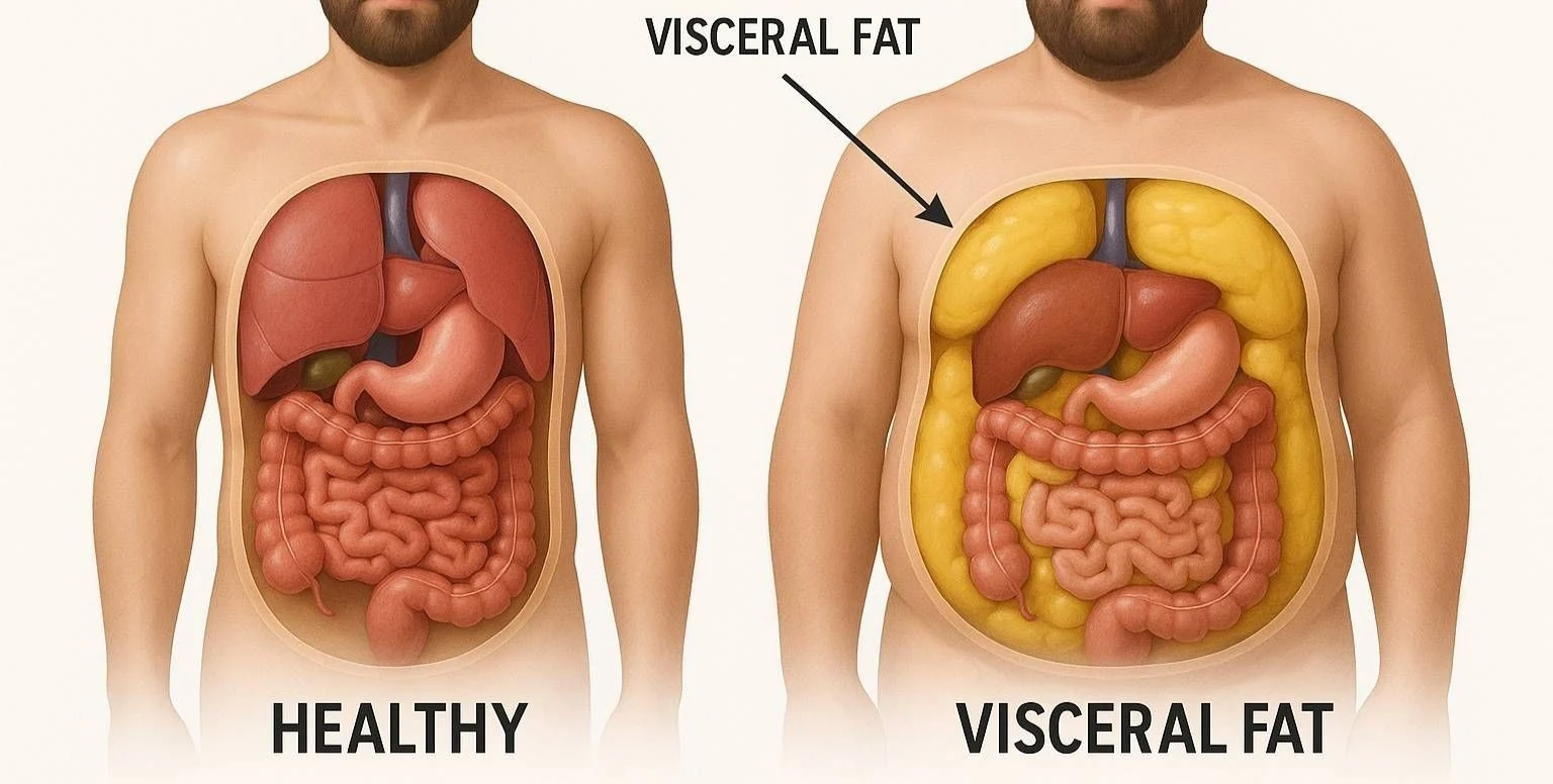
Visceral fat, the adipose tissue that accumulates around the liver, pancreas, and intestines, is one of the most clinically significant body fat compartments. Unlike subcutaneous fat (the fat under your skin), visceral fat is metabolically active. Specifically, it releases inflammatory signalling molecules, including TNF-α, IL-6, resistin, and leptin, which are linked to insulin resistance, hepatic inflammation, and cardiovascular plaque formation. Consequently, elevated visceral fat, as measured by CT or MRI, is an independent risk marker for cardiovascular and metabolic disease, including Type 2 diabetes.[5]
Furthermore, visceral fat may be a particular concern in Indian patients. In a study of 822 adults in Canada matched for BMI, people of South Asian origin had relatively more abdominal fat, and particularly more visceral fat, than people of European origin.[9] This means that an Indian patient with a BMI in the normal range may still carry a Visceral Fat Level of 10 or above, the level InBody advises staying under.[10] In contrast, a standard weighing scale or BMI measurement may classify this patient as healthy. Moreover, even a comprehensive blood panel cannot quantify visceral fat directly; it can only reflect its downstream metabolic consequences. As a result, InBody’s Visceral Fat Level (VFL) measurement is one of the most clinically useful markers the scan provides. Note that InBody describes VFL as an estimate of the fat surrounding the internal organs, not a direct imaging measurement.[10]
2. They Quantify Your True Muscle Mass, Not Just Your Weight
Skeletal muscle mass is arguably the most important and most underappreciated health metric in modern clinical practice. Specifically, skeletal muscle performs three functions that are directly relevant to metabolic disease prevention. First, it accounts for approximately 80% of insulin-stimulated glucose uptake under experimental (euglycaemic clamp) conditions.[11] In a US national survey of 13,644 adults, higher muscle mass relative to body size was associated with better insulin sensitivity and a lower prevalence of prediabetes, even after adjusting for general and central obesity.[6] Second, it is one of the main sources of glutamine, an amino acid that immune cells, including lymphocytes and macrophages, need in large amounts.[12] Third, contracting muscle releases signalling proteins called myokines that act on other organs, including fat tissue, the liver, and the immune system.[13]
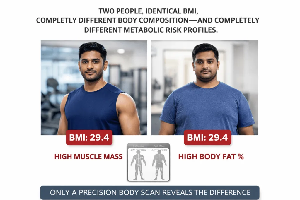
Consequently, a patient with below-norm skeletal muscle mass may face compounded risks, including poorer glucose metabolism and accelerated age-related functional decline. Moreover, these risks are invisible on a standard scale, because muscle loss is frequently masked by concurrent fat gain, meaning total body weight may remain stable even as body composition deteriorates dramatically. Therefore, InBody’s Skeletal Muscle Mass (SMM) measurement, expressed in kg and compared against age- and sex-adjusted population norms, provides information that a weighing scale or BMI cannot offer.
3. They Estimate Your Body Fat Percentage, Where Indian Thresholds Differ From Western Ones
Body fat percentage tells you what proportion of your total body mass is fat tissue. In clinical practice, this is often a more informative metric than BMI, because it estimates the fat burden directly rather than inferring it from height-to-weight ratio. Furthermore, body fat percentage allows clinicians to identify patients who are “overfat” despite appearing slim, the “normal weight obesity” phenotype that appears to be common in Indian populations. In a Singapore study of 291 adults, Indians had the highest body fat percentage at the same BMI of the three ethnic groups studied.[14]
Specifically, body fat thresholds for obesity appear to differ between populations. In a DXA study of 2,347 Indian adults, a total body fat of 25% in men and 30% in women corresponded to a BMI of about 22 kg/m², and body fat was higher in Indians than in Western populations at the same age and BMI.[15] A 2025 Indian consensus on the definition of obesity lists body-fat cut-offs of above 25.5% in men and above 38% in women.[16] Moreover, these body fat levels can occur in Indian patients at BMI values well below the standard obesity cut-off of 30. As a result, a Body Fat Percentage measurement read against these Indian reference points can help identify at-risk Indian patients who would pass a standard BMI-based health screening without any flag.
4. They Map Where Your Fat Is Located Segment by Segment
Not all fat is equally dangerous. In fact, the location of fat in the body is as clinically significant as the total fat mass. Specifically, InBody’s segmental fat analysis estimates fat in each body segment: right arm, left arm, trunk, right leg, and left leg.[17] Consequently, it can reveal an android fat distribution pattern, upper-body and trunk-dominant fat accumulation, which is associated with insulin resistance, cardiovascular disease, and metabolic syndrome, and which is common in Asian Indians, who tend to carry excess abdominal fat.[18]
Furthermore, waist circumference remains a simple screening tool for abdominal obesity in Indians, with consensus cut-offs of 90 cm in men and 80 cm in women.[18] InBody’s segmental fat analysis, particularly the trunk fat component, adds information about how fat is distributed across the trunk and limbs, although it is an estimate and does not replace a waist measurement. In addition, tracking changes in trunk fat over time, in response to dietary and exercise interventions, gives clinicians data on whether the trunk fat is responding to treatment.
5. They Detect Muscle Imbalances Between Your Left and Right Sides
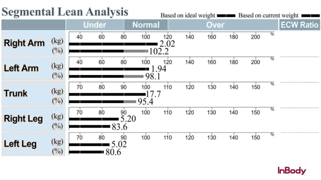
InBody’s segmental lean analysis measures skeletal muscle mass in each of the five body segments separately. This detailed approach helps identify muscle imbalances between the left and right sides, as well as between the upper and lower body. These imbalances are often missed in regular assessments.
In clinical practice, this level of detail is especially useful in three key situations.
First, for athletes and active individuals, a noticeable difference in muscle mass between the dominant and non-dominant limb can flag a muscle imbalance worth addressing in training. This is particularly important for Indian cricketers, tennis players, and runners, where one side of the body is used more intensely.
Second, for elderly patients, muscle loss in the legs is a major concern. Sarcopenia (low muscle mass together with low muscle strength or physical performance) is associated with a higher risk of falls and fractures in older adults.[19]
Third, for patients recovering from surgery or injury, segmental lean analysis helps track progress in the affected limb. It provides a clear and objective way to monitor recovery and decide when it is safe to return to normal activity.
6. They Track Body Water Balance (ECW/TBW), a Marker Linked to Inflammation and Illness
The ECW/TBW ratio (extracellular water to total body water) is a useful but often overlooked marker in preventive medicine. Specifically, this ratio reflects the distribution of body water between intracellular and extracellular compartments. Most healthy people have a ratio of around 0.380, with an acceptable range of 0.360 to 0.390.[10][17] When the ratio rises above 0.390, it may indicate swelling or excess extracellular water. The finding is non-specific: causes can include fluid retention, inflammation, poor nutritional status, or disease, so a persistently high value should prompt clinical correlation rather than a conclusion on its own.
Furthermore, a higher ECW/TBW ratio has been reported in several clinically significant conditions. In 741 centrally obese adults, a higher ratio was associated with more visceral fat and fat mass,[20] and in 193 patients on haemodialysis, a higher ratio was correlated with protein-energy wasting and independently associated with a higher risk of death.[21] Moreover, a scoping review of 36 studies concluded that bioimpedance is a suitable and valid method for assessing body composition in oncology patients under active treatment.[22] However, bioimpedance is less well validated when hydration is abnormal, so results in these groups should be interpreted by the treating clinician.[23] Consequently, this marker from an InBody precision body scan can be a useful signal across several specialities.
7. They Estimate Your Basal Metabolic Rate, a Starting Point for Nutrition Prescriptions
Basal Metabolic Rate (BMR), the number of calories your body burns at complete rest, is the foundation of any evidence-based nutrition prescription. However, population-formula BMR calculations, such as the Harris-Benedict equation, estimate BMR from age, sex, height, and weight rather than from body composition. A reanalysis of the original Harris-Benedict data found that lean body mass was the single best predictor of BMR, and that sex and age added little.[24] As a result, formula-based estimates can be less accurate for individuals whose muscle and fat proportions differ from the average, which a dietitian should keep in mind when setting a calorie prescription.
In contrast, InBody estimates BMR from measured lean body mass using a regression equation, because lean body mass is closely related to BMR.[24][25] Consequently, InBody-derived BMR reflects an individual’s body composition, although it remains an estimate and not a direct measurement of resting energy expenditure. Furthermore, tracking BMR changes over time can indicate whether a weight-loss intervention is preserving lean mass (BMR maintained or increasing) or causing lean mass loss (BMR declining), a distinction that is clinically useful but invisible on a standard scale.
8. They Track the Sarcopenia Trajectory Before It Becomes Severe
The Asian Working Group for Sarcopenia (AWGS 2019) defines sarcopenia as low muscle mass plus low muscle strength and/or low physical performance. For muscle mass measured by bioimpedance, the AWGS cut-offs for skeletal muscle mass index (SMI) are below 7.0 kg/m² in men and below 5.7 kg/m² in women (by DXA, the cut-off for women is 5.4 kg/m²).[26] It is a progressive condition that develops gradually, often well before it causes obvious functional impairment. In India, contributing factors may include low-quality dietary protein, sedentary lifestyles, and age-related muscle loss; Indian diets derive almost 60% of their protein from cereals, which have relatively low digestibility and quality.[27] Furthermore, the AWGS publishes its own cut-offs for Asian populations. These were developed mainly from studies in East Asia, and the AWGS notes that more research is needed in South Asia, so Indian-specific thresholds may differ.[28]
Therefore, InBody’s Skeletal Muscle Index measurement, available directly from the scan result sheet, can help clinicians identify patients whose muscle mass is trending towards the sarcopenia range before they meet the diagnostic threshold. Note that a low SMI alone does not diagnose sarcopenia; the AWGS definition also requires low muscle strength and/or low physical performance. In practice, this means that a 45-year-old patient who is currently borderline-low in muscle mass can be advised on protein intake and resistance training now, which may help reduce the risk of the falls, fractures, and loss of functional independence that sarcopenia is associated with in later life.[19] In addition, annual or six-monthly InBody scans allow clinicians to track the trajectory and see whether interventions are helping to maintain muscle.
9. They Provide a Single Composite Score That Tracks Your Health Over Time
The InBody Score is a composite number that summarises muscle-fat balance in a single, easily communicated figure. According to InBody, the baseline score is 80; points are added when lean mass is above, or fat mass below, the healthy average for the same height and sex, and subtracted when the opposite is true.[29] A score above 80 therefore indicates a more favourable muscle-fat balance than the healthy average. Moreover, tracking the InBody Score over serial scans at 8–12 week intervals provides an objective, motivating measure of whether a patient’s body composition is improving in response to lifestyle, dietary, or pharmacological interventions.
Furthermore, in clinical practice, the InBody Score can serve a purpose beyond data: it can act as a patient engagement tool. Specifically, a tangible, improving number may help motivate patients to stay with a diet and exercise programme. As a result, the InBody Score can help address the adherence gap that is a common barrier to preventive care.
10. They Show Whether Weight Lost on GLP-1 Medications Is Fat or Lean Tissue
GLP-1 receptor agonists, such as semaglutide, liraglutide, and their equivalents, are increasingly prescribed in India for Type 2 diabetes and obesity management. However, a review of clinical studies found that in over half of them, 20% to 50% of the weight lost on GLP-1 receptor agonists and SGLT2 inhibitors was lean body mass rather than fat,[30] and a 2026 meta-analysis of randomised trials put the lean share at about one-third (33.3%) of total weight loss for incretin-based therapies.[31] (Lean mass includes water and organs as well as muscle.) This is a clinical concern: the patient’s weight on the scale decreases, their doctor is satisfied, and meanwhile, their lean mass may be declining, which warrants attention.
Consequently, body composition scanning can be a useful way to monitor patients on GLP-1 and weight-loss pharmacotherapy. Specifically, serial scans at baseline, 3, 6, and 12 months can help clinicians distinguish fat loss from lean mass loss and see whether the scale reading is falling at the cost of lean tissue. Moreover, when a large lean mass loss is detected early, the treating clinician can review protein intake, add resistance training, and reconsider the treatment plan; reviews of these therapies recommend strategies to preserve lean mass, particularly exercise.[30][31] In other words, the InBody scan doesn’t just reveal the problem; it gives clinicians the information to act early.
All 10 Precision Body Scan Markers: Clinical Reference Table
The following table summarises all 10 health markers revealed by an InBody precision body scan, the clinical significance of each, and a reference or action threshold.
| S.No. | Marker | What It Reveals | Reference / Action Threshold | Available On |
|---|---|---|---|---|
| 1 | Visceral Fat Level (VFL) | Fat surrounding internal organs; an independent risk marker for T2DM and CVD | VFL 10 or above = above the level InBody advises staying under[10] | All models |
| 2 | Skeletal Muscle Mass (SMM) | Total lean muscle in kg vs age/sex norm; relevant to glucose disposal | Below the age/sex norm: discuss with a clinician; low muscle mass plus low strength or physical performance may indicate sarcopenia[26] | All models |
| 3 | Body Fat Percentage (PBF) | Proportion of total weight that is fat; helps distinguish true obesity from BMI-defined obesity | >25% men / >30% women in Indian DXA data[15]; a newer consensus lists >25.5% / >38%[16] | All models |
| 4 | Segmental Fat Analysis | Estimated fat mass per limb and trunk; shows android vs gynoid distribution | Higher trunk fat suggests an android (abdominal) pattern, which is associated with metabolic and cardiovascular risk | 570, 770, 970 |
| 5 | Segmental Lean Analysis | Muscle mass per limb and trunk; reveals imbalances and deficits | Marked left-right asymmetry = imbalance to address in training; low leg muscle mass = higher fall risk in older adults[19] | All models |
| 6 | ECW/TBW Ratio | Body water balance; a higher ratio can reflect oedema, inflammation or poor nutritional status | Acceptable range 0.360–0.390; above 0.390 = outside the range, discuss with a clinician[10] | 570, 770, 970 |
| 7 | Basal Metabolic Rate (BMR) | Estimated resting caloric burn from lean mass; a nutrition prescription baseline | Declining BMR over serial scans may indicate lean mass loss (for example from dietary restriction or GLP-1 therapy) | All models |
| 8 | Skeletal Muscle Index (SMI) | Height-adjusted muscle mass; one criterion in the AWGS sarcopenia definition | <7.0 kg/m² men / <5.7 kg/m² women by bioimpedance (AWGS 2019 cut-offs) | All models |
| 9 | InBody Score | Composite muscle-fat balance index; patient engagement and progress tracking | 80 = InBody baseline (healthy average); above 80 = more favourable muscle-fat balance; track the trend over 8–12 week cycles[29] | All models |
| 10 | Lean-to-Fat Change Ratio | On serial scans, helps distinguish fat loss from lean mass loss during weight loss therapy | A large share of weight lost as lean tissue may warrant a review of protein intake, exercise or the treatment plan with the clinician[30] | All models (serial scan) |
Illustrative Case: What a Precision Body Scan Could Find That 3 Years of Standard Checks Missed
The following example is illustrative: it is not a published case report, and its figures are examples chosen to show how the markers can be read together. A 44-year-old female accountant in Hyderabad had attended annual corporate health checks for three consecutive years. Each year, her results came back reassuring: BMI 23.1, fasting glucose 5.1 mmol/L (normal), cholesterol within range, blood pressure 118/76 mmHg. Consequently, she was classified as low metabolic risk and received no intervention recommendations.
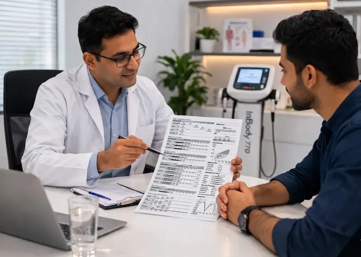
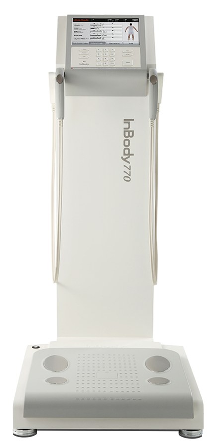
However, she reported persistent fatigue, increasing difficulty managing her weight despite no change in diet, and a family history of Type 2 diabetes on both sides. As a result, her physician requested an InBody 770 precision body composition scan. The results were clinically striking.
Specifically, the scan revealed: Visceral Fat Level 12 (high risk, 2 units above the level of 10 that InBody advises staying under), Body Fat Percentage 37.4% (above the 30% female threshold from Indian DXA data), Skeletal Muscle Mass 2.9 kg below the population norm for her age and height, and an ECW/TBW ratio of 0.392 (marginally above the 0.390 upper limit of InBody’s acceptable range). Furthermore, her InBody Score was 61, below the InBody baseline of 80. In other words, three years of standard health checks had classified her as healthy while all four body composition markers were outside their reference ranges.
Subsequently, a 16-week structured programme was initiated: a protein target of 1.2 g/kg/day, progressive resistance training three times per week, and a 500 kcal daily deficit targeting fat loss without muscle compromise. At the 16-week InBody follow-up scan, the results showed improvement across all four markers.
| VFL 12 → 8 Visceral Fat Level dropped 4 units out of high-risk zone | +2.4 kg SMM Skeletal Muscle Mass gained, with estimated BMR up by 89 kcal/day | PBF 37.4% → 31.2% Body Fat Percentage reduced by 6.2 percentage points | Score 61 → 74 InBody Score improved 13 points, closer to the 80 baseline |
In addition, her fasting glucose at the 16-week clinical review had reduced to 4.7 mmol/L, and her fatigue symptoms had improved noticeably. This case illustrates the core clinical argument for precision body scanning: standard health screening did not fail because of negligence; it failed because it was not designed to see what InBody sees.
Who Should Get a Precision Body Scan And How Often
A precision body scan using InBody technology can be useful for a broad range of patients and contexts. Specifically, the following groups may benefit most from regular scanning and are a natural focus for proactive body composition monitoring in India.
- Adults over 35 with a family history of diabetes, cardiovascular disease, or metabolic syndrome: These patients may be at higher risk from the thin-fat Indian phenotype and should be scanned annually at minimum, regardless of current BMI or blood panel status.
- Patients on GLP-1 receptor agonists or weight-loss pharmacotherapy: Serial scans at baseline, 3, 6, and 12 months can help distinguish fat loss from lean mass loss and inform decisions about the treatment plan.
- Post-COVID patients with persistent fatigue or functional impairment: ECW/TBW ratio and SMM monitoring can provide objective data on fluid balance and muscle recovery.
- Athletes and active individuals at any level: Segmental lean analysis, SMM tracking, and InBody Score monitoring can help coaches and sports medicine clinicians monitor training response and spot muscle imbalances.
- Elderly patients over 60: SMI monitoring against AWGS thresholds, segmental leg muscle analysis, and ECW/TBW ratio tracking can support sarcopenia screening and monitoring in India’s ageing population.
- Corporate employees in sedentary roles: Annual InBody scanning in workplace wellness programmes can help identify employees with raised visceral fat and metabolic risk at a stage when dietary and exercise changes may help improve the trajectory.
- Patients in oncology, nephrology, or post-surgical care: ECW/TBW ratio and serial SMM monitoring can provide objective data on fluid balance and lean mass that the treating team can consider alongside clinical findings. Bioimpedance is less well validated when hydration is abnormal, so results need careful interpretation.[23]
SUGGESTED SCANNING FREQUENCY BY CLINICAL CONTEXT (general guidance, not a formal clinical guideline)
General health monitoring (adults 35+): Every 6–12 months
Active weight management or fitness programme: Every 8–12 weeks
GLP-1 / pharmacotherapy monitoring: Baseline + every 3 months
Post-COVID recovery: Baseline + every 6–8 weeks until ECW/TBW normalises
Sarcopenia prevention (adults 60+): Every 6 months
Elite athletic performance: Every 4–6 weeks during competitive season
Oncology/nephrology nutritional monitoring: Monthly or as clinically indicated
How to Prepare for an InBody Precision Body Scan
To obtain the most reliable InBody scan results, the following preparation guidelines should be followed, in line with the manufacturer’s instructions.[25] Adhering to these conditions helps ensure that the impedance measurements reflect body composition rather than transient physiological fluctuations.
- Fast for 2–3 hours before scanning. Food and drink in the gastrointestinal tract can affect body water distribution and alter impedance readings.
- Avoid exercise before scanning, ideally for several hours. Exercise and sweating can cause transient fluid shifts that temporarily change the readings.
- Empty the bladder immediately before stepping on the device. A full bladder adds to body weight and can slightly affect the reading.
- Remove shoes, socks, and all footwear. Direct electrode contact with the soles of both feet is required for accurate current transmission through the lower body segments.
- Scan at the same time of day for all serial measurements. Body water levels vary through the day. Consistent scan timing, ideally in the morning before breakfast, reduces this variable across serial assessments.
- Remove all metal jewellery and accessories. Metal accessories may affect the measurement, so remove as many as practical.
- Women may wish to scan at the same point in the menstrual cycle for serial scans. Small fluid shifts across the cycle can change impedance and fat-free mass estimates slightly; in one study of 25 women, body fat percentage did not differ significantly across the cycle.[32]
Frequently Asked Questions
InBody DSM-BIA technology has been compared with DEXA, a widely used reference method for body composition, in published validation studies, which report close agreement for lean and fat mass, with small systematic differences and wider individual limits of agreement for body fat percentage.[1][2][3] It uses Direct Segmental Multi-frequency BIA, measuring five body segments with multiple frequencies, giving segmental body composition without radiation or long scan times.
No—an InBody body composition analyzer does not replace clinical tests. It complements standard assessments by providing additional data such as visceral fat, skeletal muscle mass, segmental analysis, and ECW/TBW ratio—metrics not available in routine pathology tests. For the most complete evaluation, clinicians combine body composition analysis with blood tests, blood pressure readings, and patient history.
InBody scanning is generally considered safe for most people, including many patients with diabetes, hypertension, and other chronic conditions. It uses a very low, painless electrical current and is non-invasive and radiation-free.[25] It should not be used by people with implanted electronic medical devices such as pacemakers,[25] and testing during pregnancy is generally not recommended. In people with cancer, kidney disease, or abnormal fluid balance, results should be interpreted by the treating clinician, because bioimpedance is less well validated when hydration is abnormal.[23]
Many consumer body fat scales use single-frequency, foot-to-foot BIA, which estimates body composition from limited measurements and population formulas. In contrast, InBody uses multi-frequency, segmental analysis across five body parts to report markers like Visceral Fat Level, ECW/TBW ratio, SMI, and segmental lean mass, giving a more detailed picture, although these remain BIA estimates and not direct imaging measurements.
InBody precision body scanners are available across hospitals, diabetes clinics, sports medicine centres, corporate wellness programs, and premium fitness facilities throughout India. Find an InBody scanner near you at inbody.in, or contact InBody India for institutional deployment in hospitals, clinics, or corporate wellness programs.
Key Takeaways
- Standard health screening in India, including weight, BMI, and blood panel, does not measure visceral fat distribution or skeletal muscle mass, two factors closely linked to metabolic risk. A precision body scan estimates both.
- An InBody scan reveals 10 clinically distinct health markers: visceral fat level, skeletal muscle mass, body fat percentage, segmental fat and lean analysis, ECW/TBW ratio, BMR, SMI, InBody score, and lean-to-fat change ratio in a single 60-second, radiation-free assessment.
- The thin-fat Indian phenotype, with normal BMI masking high visceral fat and low muscle mass, appears to be common in India and can be identified more reliably through body composition analysis than through weight or BMI alone.
- Patients on GLP-1 and weight-loss medication can benefit from serial InBody monitoring to check whether weight loss is coming from fat or from lean mass, including muscle.
- InBody DSM-BIA has shown close agreement with DEXA in published validation studies, making it a practical, non-invasive way to assess body composition at the bedside, although BIA values are estimates and can differ from DEXA in individuals.
- Suggested scanning frequencies range from monthly (oncology, nephrology) to annual (general health monitoring), with 8–12 week cycles typical for active weight management and fitness programmes.
Discover What Your Standard Health Check Has Been Missing
An InBody precision body scan takes 60 seconds. It costs a fraction of a standard health check. And it reveals the 10 health markers that your weighing scale, your BMI chart, and your blood panel have never been able to show you. That is not just a claim: body composition analysis by bioimpedance has been studied in peer-reviewed research for decades.
Find an InBody Scanner Near You
References
- Ling CH, de Craen AJ, Slagboom PE, Gunn DA, Stokkel MP, Westendorp RG, Maier AB. Accuracy of direct segmental multi-frequency bioimpedance analysis in the assessment of total body and segmental body composition in middle-aged adult population. Clin Nutr. 2011;30:610-615. PubMed · DOI
- McLester CN, Nickerson BS, Kliszczewicz BM, McLester JR. Reliability and agreement of various InBody body composition analyzers as compared to dual-energy X-ray absorptiometry in healthy men and women. J Clin Densitom. 2020;23:443-450. PubMed · DOI
- Anderson LJ, Erceg DN, Schroeder ET. Utility of multifrequency bioelectrical impedance compared with dual-energy x-ray absorptiometry for assessment of total and regional body composition varies between men and women. Nutr Res. 2012;32:479-485. PubMed · DOI
- Anjana RM, Unnikrishnan R, Deepa M, et al. Metabolic non-communicable disease health report of India: the ICMR-INDIAB national cross-sectional study (ICMR-INDIAB-17). Lancet Diabetes Endocrinol. 2023;11:474-489. PubMed · DOI
- Neeland IJ, Ross R, Després JP, et al. Visceral and ectopic fat, atherosclerosis, and cardiometabolic disease: a position statement. Lancet Diabetes Endocrinol. 2019;7:715-725. PubMed · DOI
- Srikanthan P, Karlamangla AS. Relative muscle mass is inversely associated with insulin resistance and prediabetes. Findings from the third National Health and Nutrition Examination Survey. J Clin Endocrinol Metab. 2011;96:2898-2903. PubMed · DOI
- Yajnik CS, Fall CHD, Coyaji KJ, et al. Neonatal anthropometry: the thin-fat Indian baby. The Pune Maternal Nutrition Study. Int J Obes Relat Metab Disord. 2003;27:173-180. PubMed · DOI
- WHO Expert Consultation. Appropriate body-mass index for Asian populations and its implications for policy and intervention strategies. Lancet. 2004;363:157-163. PubMed · DOI
- Lear SA, Humphries KH, Kohli S, Chockalingam A, Frohlich JJ, Birmingham CL. Visceral adipose tissue accumulation differs according to ethnic background: results of the Multicultural Community Health Assessment Trial (M-CHAT). Am J Clin Nutr. 2007;86:353-359. PubMed · DOI
- InBody Co., Ltd. InBody Results Interpretation (InBody 570, adult). Manufacturer documentation, accessed 1 October 2026, not peer-reviewed. Link
- DeFronzo RA, Tripathy D. Skeletal muscle insulin resistance is the primary defect in type 2 diabetes. Diabetes Care. 2009;32(Suppl 2):S157-S163. PubMed · DOI
- Cruzat V, Macedo Rogero M, Noel Keane K, Curi R, Newsholme P. Glutamine: metabolism and immune function, supplementation and clinical translation. Nutrients. 2018;10(11):1564. PubMed · DOI
- Pedersen BK, Febbraio MA. Muscles, exercise and obesity: skeletal muscle as a secretory organ. Nat Rev Endocrinol. 2012;8:457-465. PubMed · DOI
- Deurenberg-Yap M, Schmidt G, van Staveren WA, Deurenberg P. The paradox of low body mass index and high body fat percentage among Chinese, Malays and Indians in Singapore. Int J Obes Relat Metab Disord. 2000;24:1011-1017. PubMed · DOI
- Marwaha RK, Tandon N, Garg MK, Narang A, Mehan N, Bhadra K. Normative data of body fat mass and its distribution as assessed by DXA in Indian adult population. J Clin Densitom. 2014;17:136-142. PubMed · DOI
- Misra A, Vikram NK, Ghosh A, Ranjan P, Gulati S; India Obesity Commission. Revised definition of obesity in Asian Indians living in India. Diabetes Metab Syndr. 2025;19:102989. PubMed · DOI
- InBody. The Professional’s Guide to the InBody Result Sheet (e-book). InBody Co., Ltd. Manufacturer documentation, undated PDF hosted by InBody UK, not peer-reviewed. Link
- Misra A, Chowbey P, Makkar BM, et al. Consensus statement for diagnosis of obesity, abdominal obesity and the metabolic syndrome for Asian Indians and recommendations for physical activity, medical and surgical management. J Assoc Physicians India. 2009;57:163-170. PubMed
- Yeung SSY, Reijnierse EM, Pham VK, et al. Sarcopenia and its association with falls and fractures in older adults: a systematic review and meta-analysis. J Cachexia Sarcopenia Muscle. 2019;10:485-500. PubMed · DOI
- Harimawan AIW, Prabandari AASM, Wihandani DM, et al. Association between phase angle and ECW/TBW ratio with body composition in individuals with central obesity: a cross-sectional study. Front Nutr. 2025;12:1638075. PubMed · DOI
- Yajima T, Yajima K. Association of extracellular water/total body water ratio with protein-energy wasting and mortality in patients on hemodialysis. Sci Rep. 2023;13:14257. PubMed · DOI
- Branco MG, Mateus C, Capelas ML, et al. Bioelectrical impedance analysis (BIA) for the assessment of body composition in oncology: a scoping review. Nutrients. 2023;15(22):4792. PubMed · DOI
- Kyle UG, Bosaeus I, De Lorenzo AD, et al. Bioelectrical impedance analysis-part II: utilization in clinical practice. Clin Nutr. 2004;23:1430-1453. PubMed · DOI
- Cunningham JJ. A reanalysis of the factors influencing basal metabolic rate in normal adults. Am J Clin Nutr. 1980;33:2372-2374. PubMed · DOI
- InBody Co., Ltd. InBody230 User’s Manual. Manufacturer documentation, accessed 1 October 2026, not peer-reviewed. Link
- Chen LK, Woo J, Assantachai P, et al. Asian Working Group for Sarcopenia: 2019 consensus update on sarcopenia diagnosis and treatment. J Am Med Dir Assoc. 2020;21:300-307.e2. PubMed · DOI
- Swaminathan S, Vaz M, Kurpad AV. Protein intakes in India. Br J Nutr. 2012;108(Suppl 2):S50-S58. PubMed · DOI
- Chen LK, Liu LK, Woo J, et al. Sarcopenia in Asia: consensus report of the Asian Working Group for Sarcopenia. J Am Med Dir Assoc. 2014;15:95-101. PubMed · DOI
- InBody USA. How is InBody Score calculated? LookinBody FAQ. Manufacturer documentation, accessed 1 October 2026, not peer-reviewed. Link
- Sargeant JA, Henson J, King JA, Yates T, Khunti K, Davies MJ. A review of the effects of glucagon-like peptide-1 receptor agonists and sodium-glucose cotransporter 2 inhibitors on lean body mass in humans. Endocrinol Metab (Seoul). 2019;34:247-262. PubMed · DOI
- Busk-Cirera L, Carlsen IØ, Madsen LB, et al. Effects of incretin-based therapies, diet and exercise interventions, and bariatric surgery on fat-free mass in adults with overweight or obesity: a systematic review and meta-analysis. Diabetes Obes Metab. 2026;28:7737-7751. PubMed · DOI
- Gleichauf CN, Roe DA. The menstrual cycle’s effect on the reliability of bioimpedance measurements for assessing body composition. Am J Clin Nutr. 1989;50:903-907. PubMed · DOI

Most trending articles
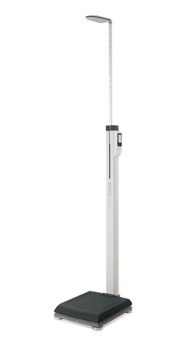
Student Fitness Screening in India: A Guide for Schools
CBSE HPE, Fit India and Khelo India rely on BMI. How schools and colleges can add body composition screening safely, with child caveats and DPDP consent.
Creatine and Your Body Composition Scan: What Changes
Creatine raises body water and lean mass readings within a week on BIA and DXA. What the studies show and how to time your scans to read progress honestly.
Fitness Testing and Body Composition for Sports Academies
A practical fitness testing battery for Indian sports academies: body composition, grip, jumps, sprints, Yo-Yo, Khelo India context and youth maturation.