
Body Composition 11 min read
Osteoporosis in India: Why Declining Muscle Mass Is the Warning Sign Before the Fracture
18% of Indian adults have osteoporosis, and 69% of over-50s have low bone density, mostly silent. See how muscle mass flags the risk early, for men and women.
Reading about body composition? Find an InBody test centre near you →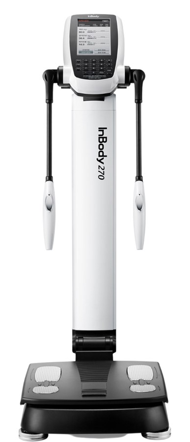
The Fracture That Comes as a Complete Surprise (It Shouldn’t)
It follows a familiar shape at orthopaedic clinics across urban India. A 58-year-old steps off a footpath awkwardly, or slips getting out of the shower, and ends up with a wrist or hip fracture wildly disproportionate to the fall itself. The scan that follows reveals osteoporosis — bone density low enough that a minor stumble was enough to break a bone. The family is stunned. This person walked daily, ate reasonably, wasn’t visibly frail. “She seemed healthy,” is the refrain doctors hear constantly.
What almost never comes up is that a warning sign had likely been visible for years — just not in the place anyone was looking. Osteoporosis and declining skeletal muscle mass are linked at a real biological level, mechanically and hormonally, closely enough that researchers use a specific term — osteosarcopenia — for the overlap. Muscle mass is far easier to track over time than bone density is, and a steady muscle-loss trend on repeat body composition tests is often the earliest visible signal that bone density is eroding alongside it — in men as well as women.
This isn’t the same story as menopause-driven fat gain, and it isn’t a general “muscle loss with age” story either. It’s about what happens when muscle and bone decline together, why that combination is dangerous, and why watching one can help catch trouble in the other before a fracture forces the issue.
Why Muscle and Bone Decline Together: The Science of Osteosarcopenia
Bone and muscle are usually discussed as separate systems — one measured with a DEXA scan, the other with a body composition test — but physiologically they are closely linked, communicating through two connected pathways.
Mechanical loading: bone responds to the pull of muscle
Bone is living tissue that remodels itself in response to mechanical stress. Most of that stress doesn’t come from body weight alone — it comes from skeletal muscle contracting against bone at every joint, step, and lift. Strong, well-loaded muscle keeps bone under a stimulus that signals it to maintain density. When muscle mass declines — from inactivity, illness, or age — that loading signal weakens, and bone has less reason to maintain itself.
Myokines and osteokines: two tissues that talk to each other
Beyond mechanics, muscle and bone signal each other biochemically. Skeletal muscle secretes hormone-like proteins called myokines — including irisin, myostatin, IGF-1, and interleukin-6 — during contraction and routine metabolism. Bone secretes its own signalling proteins, called osteokines, including osteocalcin and sclerostin. Research on this “muscle-bone crosstalk” shows several myokines directly influence bone-forming and bone-resorbing cells, while osteokines influence muscle metabolism in return — a genuine two-way hormonal conversation.
Shared risk factors accelerate both at once
The overlap deepens because muscle and bone deteriorate in response to many of the same drivers: declining estrogen or testosterone with age, inadequate protein and calcium, vitamin D deficiency, inactivity, and chronic inflammation all damage both tissues independently. This is why researchers coined the term osteosarcopenia, for the high-risk subset of people in whom sarcopenia and osteopenia/osteoporosis occur together, compounding each other’s damage.
The consequence is measurable. One study following postmenopausal women over 12 months found those with low appendicular muscle mass had significant femoral neck bone density decline over that period, while women with normal muscle mass showed no significant bone loss — evidence a muscle-mass deficit can precede and predict bone loss, not simply run alongside it. Reviews estimate the osteosarcopenia overlap affects 5–37% of community-dwelling older adults, rising to around 46% in people who’ve already had a low-trauma fracture.
Osteoporosis in India: Common, Serious, and Rarely Diagnosed Before a Fracture
Osteoporosis is not a rare condition in India — it’s widespread and poorly tracked. A large study of 31,238 Indian adults found 18.3% had osteoporosis and another 49.9% had osteopenia, the lower-density stage that precedes it — close to two in three adults with some degree of bone loss. Prevalence climbs with age: osteoporosis was found in 33.1% of postmenopausal women and 37.0% of adults 60 and above, versus 12.5% of adults under 60.
Men are affected too, more than most assume. Research in the Indian Journal of Orthopaedics estimates that roughly 50% of women and 20% of men over 50 in India will have an osteoporosis-related fracture in their remaining lifetime, and more than a third of all hip fractures nationally occur in men — undercutting the assumption that bone health is purely a menopause issue.
The bigger problem is the silence around it. A study of 455 adults aged 50–89 in Chandigarh found 69% had low bone density — osteopenia or osteoporosis combined — yet only 7.3% had ever reported a bone-related symptom to a doctor. Osteoporosis is often called a “silent disease” for exactly this reason: no pain, no visible sign, nothing until a bone breaks. In a country where DEXA screening is far less routine than a blood pressure check — typically ordered only after a fracture, rather than as part of a standard check after 40 or 50 — that silence tends to go unbroken for years.
The stakes are real. A widely cited estimate places India’s annual burden of osteoporotic hip fractures at roughly 600,000 cases — a dated 2004 projection, but still the most referenced figure in Indian orthopaedic literature. Indian studies have also found hip fractures occur at a younger age here than in Western populations, and that difficulty rising unassisted from a chair — a marker of lower-body muscle weakness — was linked to a more than 14-fold higher hip fracture risk in a Delhi case-control study. Falls themselves are common: close to a third of older Indian adults fall at least once a year, and roughly 1 in 8 of those falls results in a fracture.
Why Skeletal Muscle Mass Is the Early-Warning System Most Indians Actually Have Access To
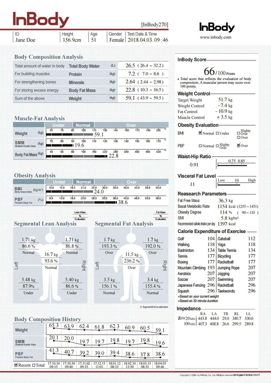
Here is the honest, important distinction: an InBody test does not measure bone density. That requires a DEXA (dual-energy X-ray absorptiometry) scan, interpreted by a doctor. InBody testing measures Skeletal Muscle Mass (SMM), body fat, and water distribution — not bone.
What InBody testing offers is something most Indians can access far more easily than a DEXA scan: a fast, repeatable read on skeletal muscle mass, taken in under five minutes at gyms, clinics, and hospitals, without a referral. Because muscle and bone decline together, a clear downward trend in Skeletal Muscle Mass across repeat InBody tests is a reasonable, evidence-supported reason to ask a doctor about a bone-density scan — well before a fracture forces that conversation.
| InBody body composition test | DEXA bone density scan | |
|---|---|---|
| What it measures | Skeletal muscle mass, body fat, water distribution | Bone mineral density (T-score) |
| Time required | Under 5 minutes | 10–20 minutes |
| Typical access | Gyms, clinics, hospitals — no referral needed | Radiology/diagnostic centres, usually needs a doctor’s order |
| Role here | Early-warning trend — flags who should ask for a bone scan | Diagnostic confirmation of osteopenia/osteoporosis |
This applies as much to men as to women. Because bone-health messaging in India is so heavily anchored to menopause, men with declining Skeletal Muscle Mass often have no equivalent trigger to think about their bones — despite carrying roughly a third of the country’s hip fracture burden, and facing their own age-related testosterone decline, which independently affects muscle and bone (covered in this blog’s piece on testosterone and muscle loss in Indian men). A downward SMM trend across consecutive InBody tests — particularly after 45, alongside reduced grip strength or difficulty rising from a low chair — is the signal worth acting on, for men and women alike.
None of this replaces a DEXA scan. It solves an access problem: DEXA screening in India isn’t yet routine, so most people never get one until something has gone wrong. Muscle-mass tracking is the more available signal that can prompt the right people to ask for that scan sooner. For a fuller method comparison, see InBody vs DEXA vs skinfold calipers.
A Bone-and-Muscle Protocol: What to Actually Do
- Do resistance training that loads bone, not just muscle. The LIFTMOR trial, published in the Journal of Bone and Mineral Research, found postmenopausal women with low bone mass doing twice-weekly high-intensity resistance and impact training — heavy compound lifts plus jumping-type loading — gained significantly more spine and hip bone density than a lighter home-exercise group, safely, challenging the assumption that weak bones mean avoiding heavy lifting. Progressively loaded compound training — squats, deadlifts, presses, rows — under supervision is the single most evidence-backed intervention for both muscle and bone.
- Get enough protein, spread across the day. Muscle protein synthesis needs a steady supply, not one large serving at dinner. Build meals around dal, paneer, curd, eggs, fish, or chicken at each sitting to support the muscle maintenance that keeps bone-loading mechanics intact.
- Treat calcium and vitamin D as a pair. Calcium-rich foods (dairy, ragi, sesame seeds, dark leafy greens) supply the raw material for bone; vitamin D lets the body absorb it. Deficiency is common in urban India despite abundant sunshine — a South Indian study found 53% of healthy men over 50 were deficient — so confirm with a blood test rather than assuming sun exposure covers it.
- Ask for a DEXA scan at the right trigger points — not only after a fracture. Reasonable prompts: any postmenopausal woman, any man or woman over 65, a family history of osteoporosis or fracture, a previous low-trauma fracture, or a sustained downward SMM trend on repeat InBody tests.
- Address fall risk directly, not just bone density. Since most fractures start with a fall, improving balance and lower-body strength (chair squats, step-ups, single-leg balance work), fixing poor home lighting and loose rugs, and getting vision checked regularly are all legitimate parts of a bone-health protocol.
- Track Skeletal Muscle Mass on a consistent cadence. Test every 3–4 months during active intervention — new training, post-fracture recovery, or the years around menopause — and roughly every 6–12 months for general monitoring after 45, to catch a declining trend before it becomes a fracture story.
Frequently Asked Questions
Can declining muscle mass really predict weak bones?
Yes, to a meaningful degree. Muscle and bone share mechanical loading and hormonal signalling pathways, so they tend to decline together. One study found postmenopausal women with low appendicular muscle mass had significant hip bone density loss over 12 months, while those with normal muscle mass did not. A sustained drop in Skeletal Muscle Mass on repeat tests is a reasonable prompt to ask about bone density testing, not a diagnosis on its own.
What is osteosarcopenia?
Osteosarcopenia is the term for having both sarcopenia (age-related muscle loss) and osteopenia or osteoporosis (reduced bone density) at once. Because muscle and bone influence each other mechanically and hormonally, the two conditions frequently occur together and compound each other’s fracture risk. Research estimates the overlap affects roughly 5–37% of community-dwelling older adults, rising sharply among those who’ve already suffered a fracture.
Does an InBody test measure bone density?
No. InBody testing measures Skeletal Muscle Mass, body fat, and water distribution — not bone mineral density, which only a DEXA scan can confirm. What InBody testing can do is flag a declining muscle-mass trend, which research links to bone density loss, giving you a practical reason to seek a DEXA scan proactively rather than waiting for a fracture.
Is osteoporosis only a concern for women after menopause?
No. Postmenopausal women carry the highest individual risk due to the drop in estrogen, but Indian research estimates roughly 20% of men over 50 will have an osteoporosis-related fracture in their remaining lifetime, and over a third of all hip fractures in India occur in men. Declining testosterone affects bone density much like declining estrogen does, so men’s bone health deserves equal attention.
At what age should I consider a DEXA bone density scan in India?
Common triggers: all postmenopausal women, all men and women over 65, a family history of osteoporosis or a previous low-trauma fracture, and a sustained decline in Skeletal Muscle Mass across repeat body composition tests. There’s no single universal age cutoff — a doctor should weigh your specific risk factors — but these are the most widely used prompts.
How often should I test my body composition to watch for this?
For most adults over 45 without an active concern, testing Skeletal Muscle Mass roughly every 6–12 months is enough to catch a meaningful downward trend. If you’re actively intervening — starting resistance training, recovering from a fracture, or navigating menopause — testing every 3–4 months gives clearer, faster feedback on whether your bone-loading capacity is moving the right way.
Know Your Muscle Trend Before a Fracture Forces the Conversation
Osteoporosis in India is common, serious, and overwhelmingly silent until a fracture makes it impossible to ignore. You cannot see bone density loss from the outside, and most people never get a DEXA scan unless something prompts them to ask for one. Declining Skeletal Muscle Mass is a real, research-backed early signal worth watching — for men well into their fifties and sixties, not only women navigating menopause — and far more accessible to track than bone density itself.
Get your Skeletal Muscle Mass baseline at inbody.in/inbody-test.php and re-test on a regular cadence so you can see the trend, not just a single number. If it’s declining, take that trend to your doctor and ask specifically about a DEXA bone density scan — a conversation far better had before a fall than after one. Related reading: how sarcopenia accelerates muscle loss in Indian adults after 35, the body composition guide to staying strong after 60, and how menopause reshapes body composition in Indian women.

Most trending articles
वज़न बढ़ाने के लिए क्या खाएं? सही डाइट प्लान (सिर्फ ज़्यादा खाना नहीं)
सिर्फ ज़्यादा खाने से मांसपेशी नहीं बनती, चर्बी बढ़ सकती है। सही कैलोरी सरप्लस, प्रोटीन और डाइट प्लान जो वज़न को सही तरीके से बढ़ाए — पूरी जानकारी हिंदी में।
भारत में प्रोटीन कितना ज़रूरी है? असली आंकड़े और सही फॉर्मूला
IMRB के एक सर्वे में लगभग 73% भारतीय आहार प्रोटीन-कम पाए गए। सही प्रोटीन टारगेट कैसे निकालें और किन फूड्स से पूरा करें — पूरी जानकारी हिंदी में।
Metabolic Age क्या होती है? आपकी उम्र से ज़्यादा है या कम?
आपकी असली उम्र 34 है पर शरीर 50 साल जैसा व्यवहार कर सकता है। Metabolic age क्या है, मांसपेशी घटने के जोखिम क्या हैं और इसे कैसे घटाएं — पूरी जानकारी।